He Described Microbial Cells Using a Compound Microscope
Cells were grown in Dulbeccos Modified Eagle Medium DMEM supplemented with 10 fetal bovine serum FBS Glutamax and antibiotic-antimycotic Thermo Fisher Scientific in a humidified incubator at 5 CO 2 and 37C under. Viable cells PI cells were analysed at a constant flow rate and calculated as a percentage of control cells treated with vehicle using the.

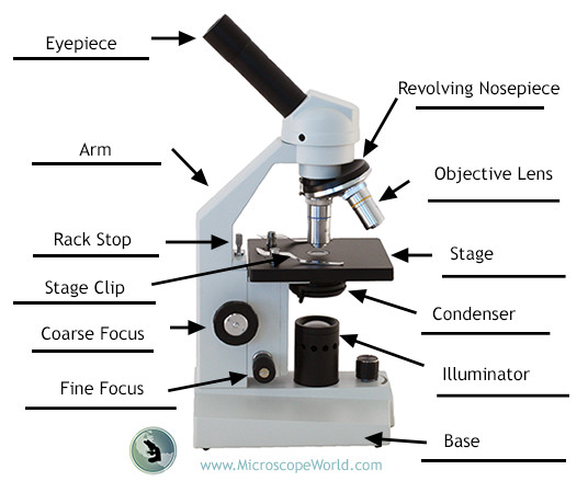
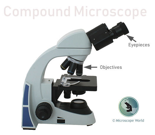
Compound Microscope Definition Labeled Diagram Principle Parts Uses
Activation of the ERK pathway has been observed in cells infected with a number of HCoVs including SARS-CoV MERS-CoV and HCoV-229E.

. Microscopy is the technical field of using microscopes to view objects and areas of objects that cannot be seen with the naked eye objects that are not within the resolution range of the normal eye. Extracellular signal-regulated kinase 12 ERK12 ERK5 c-Jun N-terminal kinase JNK and the p38 group of protein kinases. Optical electron and scanning probe microscopy along with the emerging field of X-ray microscopy.
Four distinct subgroups within the MAP kinase family have been described. Isolation - Using techniques that allow for the growth of individual colonies that can be further investigated Inspection - Observation of microbial growth characteristics with the naked eye or a microscope Identification - Using additional tests to determine the specific identity or a microbe including genetic immunological or phenotypic. In prokaryotes the primary function of the cell wall is to protect the cell from internal turgor pressure caused by the much higher concentrations of proteins and other molecules inside the cell compared to its external.
There are three well-known branches of microscopy. Neonatal primary dermal fibroblasts WT and Tlr2 were isolated by our laboratory as previously described. The cell envelope is composed of the cell membrane and the cell wallAs in other organisms the bacterial cell wall provides structural integrity to the cell.
Using bone marrow transplantation and cell type-specific Casp3 knockout mice we demonstrated that Casp3GSDME-mediated pyroptosis in renal parenchymal cells but not in hematopoietic cells.

What Is A Compound Microscope Microscope World Blog

Cell Morphology Under Compound Microscope Download Scientific Diagram
No comments for "He Described Microbial Cells Using a Compound Microscope"
Post a Comment